2021/10/18 | Research | Surgical technologies
Temporal bone properties inform robotic ear surgery
Quantitative assessment of bone density and thickness in computed-tomography images offers great potential for preoperative planning procedures in ear surgery. Together with the Departments of ENT and Neuroradiology Inselspital, the ARTORG Center has applied quantitative CT imaging to propose novel preoperative indices to identify optimal regions for screw placement and effective implant positioning.
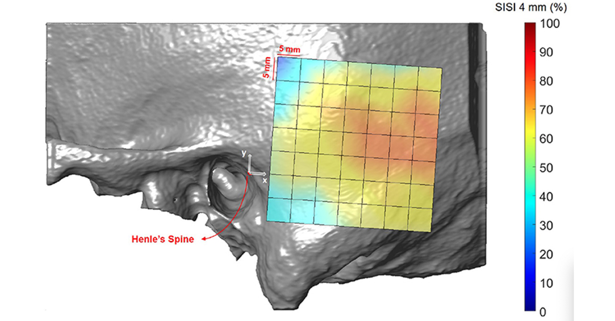 Visualization of the screw implantation safety index (SISI) for a screw length of 4 mm in the region of interest, averaged across all subjects.
(https://doi.org/10.3389/fsurg.2021.740008)
Visualization of the screw implantation safety index (SISI) for a screw length of 4 mm in the region of interest, averaged across all subjects.
(https://doi.org/10.3389/fsurg.2021.740008)
The team retrospectively analyzed CT scans of subjects undergoing cochlear implantation as well as scans of ex-vivo specimens. To assess temporal bone thickness and cortical bone density the researchers used an automated algorithm referenced by an anatomy-based coordinate system. Two indices were proposed to include information of both bone density and bone thickness for the preoperative assessment of safe screw positions (Screw Implantation Safety Index, SISI) and mass distribution (Column Density Index, CODI). In addition, the effects of age, gender, ear side and position on bone thickness, cortical bone density and the distribution of the indices were assessed.
The proposed preoperative indices (SISI and CODI) can be applied to patient-specific cases to identify optimal regions with respect to bone density and thickness for safe screw placement and effective implant positioning. The approach can thus provide quantitative information about temporal bone density and thickness for applications in robotic and computer-assisted ear surgery.