2021/12/14 | Research | Virtual & Augmented Reality
VR vs. conventional visual field testing in glaucoma
Perimetry testing for glaucoma diagnosis and progression management is currently performed with specialized setups in clinics. The Artificial Intelligence in Medical Imaging lab (AIMI) has now investigated if a VR-based perimetry test could bring comparable benefits as traditional testing while being more mobile (independent of hospital infrastructure).
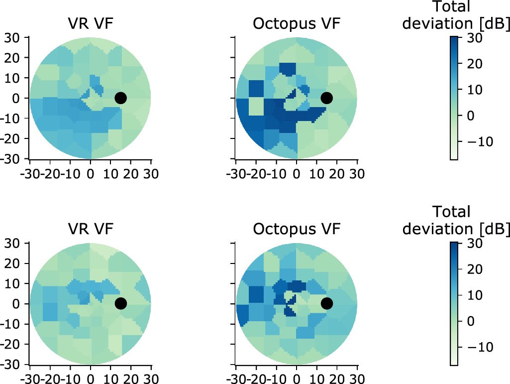 Two glaucomatous visual field examples. Each row compares acquisitions by VR perimetry (left) and by Octopus 900 (right). The black circle corresponds to the blind spot. Blue colors reflect higher deviations, that is, deeper defects.
(https://doi.org/10.1167/tvst.10.12.10)
Two glaucomatous visual field examples. Each row compares acquisitions by VR perimetry (left) and by Octopus 900 (right). The black circle corresponds to the blind spot. Blue colors reflect higher deviations, that is, deeper defects.
(https://doi.org/10.1167/tvst.10.12.10)
In collaboration with the Department of Ophthalmology of the Inselspital, researchers investigated how well a virtual reality (VR) perimetry system evaluated the visual fields of both healthy subjects and glaucoma patients when compared to a clinically and routinely used perimeter. A prospective, year-long, single center study was conducted at the Inselspital with 70 subjects (36 healthy, 34 glaucoma with early to moderate visual field loss). The authors concluded that a clinically used perimeter and the proposed VR perimetry system have comparable performances in the studied setting, suggesting that VR perimeters have the potential to assess VFs with high enough confidence, while rendering visual field tests portable and thus more accessible.
In a previous study, the team had investigated how a patented AI-based perimetry testing (sequentially optimized reconstruction strategy, SORS) performed in visual field examinations for glaucoma patients as compared to current testing schemas. The 12-month clinical study had demonstrated that the novel perimetry strategy SORS could be used in routine clinical practice with comparable utility to the current standard with the benefit of providing a shorter (i.e. 70% less time) and more comfortable perimetry experience for patients.