2022/08/23 | Research | Biomechanics
New insights into Otosclerosis with microCT
The Image Guided Therapy (IGT) lab in collaboration with the EMPA and the Departments of ENT and Neuroradiology of the Inselspital have for the first time analyzed an otosclerotic cochlea using microCT technology. The higher resolution as compared to standard CT imaging usually employed in Otosclerosis diagnosis revealed strong tissue remodeling activity even in early disease stages, previously invisible.
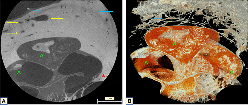 MicroCT of the cochlea apex (2.48um). A- Neovascularization of the otospongiotic bone (yellow arrows). B- mesh reconstruction of the apex with a Carp-head-shaped cochleolith and fibrotic tissue.
(https://doi.org/10.3389/fradi.2022.965474)
MicroCT of the cochlea apex (2.48um). A- Neovascularization of the otospongiotic bone (yellow arrows). B- mesh reconstruction of the apex with a Carp-head-shaped cochleolith and fibrotic tissue.
(https://doi.org/10.3389/fradi.2022.965474)
Otospongiotic plaques can be seen on conventional computed tomography (CT) as focal lesions around the cochlea. However, the resolution remains insufficient to enable evaluation of intracochlear damage. MicroCT technology provides resolution at the single micron level, offering an exceptional amplified view of the otosclerotic cochlea. In this publication, a group of engineers, physicists, and MDs for the first time present a radiological and a three-dimensional (3D) anatomical study of an otosclerotic cochlea using microCT. 3D-segmentation of the human cochlea was performed, providing an unprecedented view of the diseased area without the need for decalcification, sectioning, or staining.
Given the recently described importance of the osseous spiral lamina in hearing mechanics, the structural anatomical alterations of the otosclerotic cochlea presented here, will have a great impact on further treatment of patients presenting with sensorineural hearing loss