2022/02/08 | Research | Biomechanics
Quantifying ureteral stent encrustations
Location and extent of encrustation in ureteral stents for different diseases may be informative for patient management and the development of newer stent generations. The Urogenital Engineering Lab at ARTORG has investigated stent encrustation patterns from stone and kidney transplant patients in collaboration with the Inselspital and the Cantonal Hospital Olten.
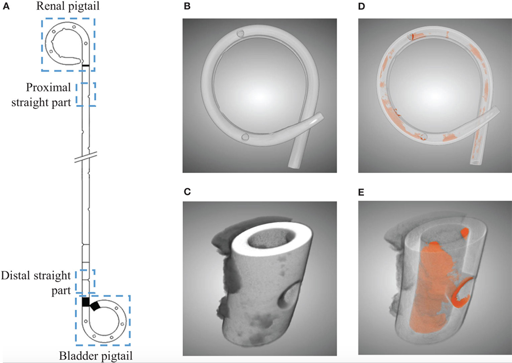 Illustration of the four separated sections from a ureteral stent (A). Three dimensional µ-CT images from the pigtail and straight part before (B, C) and after (D, E) segmentations, respectively. Luminal encrustations from the segmentation results are shown in orange, where stents are rendered semi-transparent for better visualization.
(https://doi.org/10.3389/fruro.2022.836563)
Illustration of the four separated sections from a ureteral stent (A). Three dimensional µ-CT images from the pigtail and straight part before (B, C) and after (D, E) segmentations, respectively. Luminal encrustations from the segmentation results are shown in orange, where stents are rendered semi-transparent for better visualization.
(https://doi.org/10.3389/fruro.2022.836563)
Twenty-seven double-J ureteral stents were collected from patients with stone disease or who underwent kidney transplantation. Encrustations on stent samples were quantified by means of micro−Computed Tomography and semantic segmentation using a Convolutional Neural Network model.
The quantitative results of the study suggest side holes as primary sites for initial encrustations. In both patient groups, stent pigtails were more susceptible to encrustations. Stone patients showed higher encrustation levels over prolonged indwelling times. The study team plans to clarify reasons for the different encrustation patterns found in stents placed for stone and transplant patients in upcoming experimental and numerical results produced at the ARTORG Center.