2023/03/07 | Research | Biomechanics
3D femur reconstruction from 2D DXA scans
Osteoporosis is currently diagnosed based on areal bone mineral density (aBMD) computed from 2D DXA scans. But aBMD does not take femoral geometry and density distribution into account. The Musculoskeletal Biomechanics (MSB) lab has now evaluated, in collaboration with the Department of Osteoporosis at the Inselspital, a 2D-3D DXA reconstruction software and compared femoral strength computed from the reconstructed 3D DXA and from QCT scans.
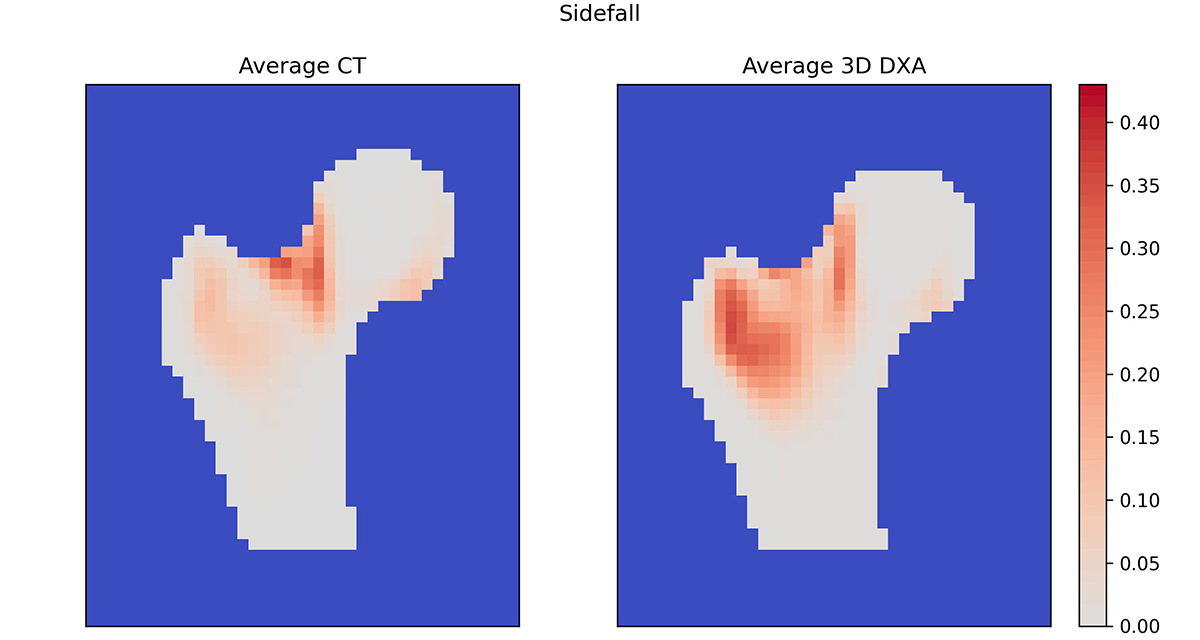 Average damage maps for QCT and 3D DXA mapped on the standard femur.
(https://doi.org/10.3389/fbioe.2023.1111020)
Average damage maps for QCT and 3D DXA mapped on the standard femur.
(https://doi.org/10.3389/fbioe.2023.1111020)
Quantitative Computed Tomography (QCT) combined with finite element (FE) analysis can deliver improved femoral strength predictions but is not widely used because of high costs and the radiation implied. As an alternative to QCT, the ARTORG MSB team evaluated the 3D-Shaper software (3D-Shaper Medical, Spain) to reconstruct the 3D shape and density distribution of the femur from 2D DXA scans. Results showed that the 3D-Shaper generates an altered BMD distribution compared to QCT but, after careful density calibration, shows an interesting potential for deriving a standardized femoral strength from a standard DXA scan.
Link to the study: https://www.frontiersin.org/articles/10.3389/fbioe.2023.1111020/full
Musculoskeletal Biomechanics