2023/09/19 | Research | Artificial Intelligence
AI-assisted brain tumor measurements
Automated tumor segmentation for glioblastoma is promising, yet little is known about longitudinal accuracy of automated measurements to assess treatment response. The Medical Image Analysis lab compared assessment consistency by two AI-based tumor segmentation tools against expert ratings over time, identifying a need for further improvement.
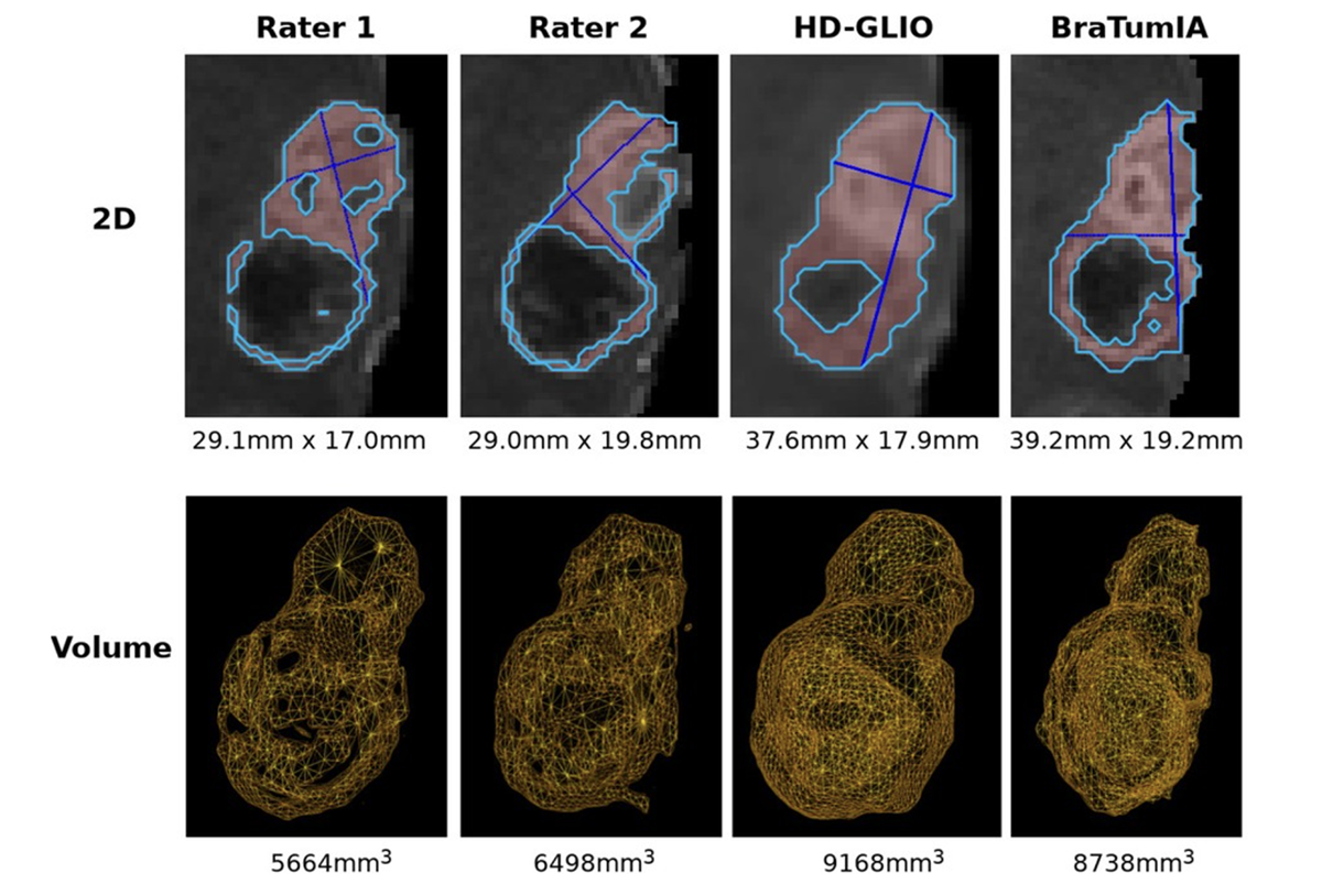 Examples of 2D and volumetric contrast-enhanced quantification. The basis for all two-dimensional quantification is a segmentation (by a human expert or automated segmentation tool) calculated automatically.
(https://doi.org/10.3389/fradi.2023.1211859)
Examples of 2D and volumetric contrast-enhanced quantification. The basis for all two-dimensional quantification is a segmentation (by a human expert or automated segmentation tool) calculated automatically.
(https://doi.org/10.3389/fradi.2023.1211859)
The study investigated performance by BraTUmIA and HD-GLIO for automated response assessment (including longitudinal segmentation and consistent tumor measurement) on the single-center retrospective dataset LUMIERE.
The tumor volume trend agreement calculated between segmentation volumes and the expert bi-dimensional measurements was high (HD-GLIO: 81.1%, BraTumIA: 79.7%). Yet both tools failed at an accurate lesion count across time.
The authors conclude that expert supervision and manual corrections are still necessary when applying the tested automated segmentation tools for automated response assessment. The longitudinal consistency of current segmentation tools needs further improvement, and evaluated ong multi-center datasets.